Retinal Technology
At Mississauga Meadowvale Optometry our only goal is to keep your eyes as healthy as possible. One of the essential tools we use to do so is retinal scanning, to detect any early signs of eye damage and disease. The sooner we detect a retinal issue, the sooner we can treat the problem. We always hope to find strong, healthy eyes but for individuals with weaker eyes and vision, we hope to improve the quality of their retinas so patients can experience optimal vision and improvement over time.

OCT-A: Our office is the first office in the GTA to offer Optical Coherence Tomography Angiography retinal imaging.
OCT provides anatomical images, while OCT-A provides additional functional information about blood flow and vascular changes that can be valuable for detecting and managing retinal diseases.
Optical Coherence Tomography (OCT)
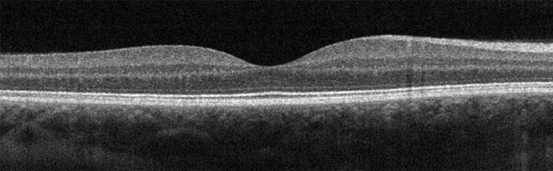 An Optical Coherence Tomography (OCT) test is an imaging test that uses light waves to take cross-section pictures of your retina, the light-sensitive tissue lining the back of the eye. With OCT, each of the retina’s distinctive layers can be seen, allowing your Doctor to map and measure their thickness. These measurements help with early detection, diagnosis and treatment guidance for retinal diseases and conditions, including age-related macular degeneration, diabetic eye disease and glaucoma to name just a few.

Heidelberg Retina Tomography (HRT)
A Heidelberg Retina Tomograph (HRT) test uses a laser light which scans the retina starting above the retinal surface capturing parallel images at increasing depths. The images are then combined to create a 3D topographic image of the optic nerve and the retinal nerve fiber layer. The HRT then uses this data to align the images and then computes a 3D topographic map of the retinal surface which is analyzed for signs of progressive damage due to glaucoma.
What is an OPTOS? What is it used for?
An OPTOS is a scanning laser technology that aids practitioners diagnosing, analyzing, documenting and monitoring pathology that may first present itself in the periphery. It is a 360° peripheral retinal scan. Without the OPTOS, pathology in the periphery may go undetected using the traditional equipment, an optomap is a screening tool for early detection of disease in the posterior segment of the eye in the context of preventative medicine, eye health and wellness. These images discover more and treat more patient diseases and pathologies more effectively. The doctor recommends having this scan done when needed.


Fundus Photograph
A Fundus Photograph is a good general retinal scan. It is taken using a retinal camera and it is used to look at different parts of the back of the eye including the retina, blood vessels, macula and the optic disc. Fundus photography allows Dr. Ahmed to detect or monitor any abnormalities related to diseases that can affect your eyes. It is considered medically necessary for conditions such as macular degeneration, retinal neoplasms, choroid disturbances and diabetic retinopathy, or to identify glaucoma, multiples sclerosis and other central nervous system abnormalities. This test is strongly recommended annually for all the patients and more frequently, if needed.
Visual Field Test
A visual field test measures a patient’s entire scope of vision (central and peripheral) by mapping the visual fields of each eye individually. Certain populations are at a higher risk for damage to the optic nerve due to glaucoma. Visual field testing is most often used to detect any signs of this damage. In addition, it is also useful for detecting central or peripheral retinal disease, eyelid conditions, optic nerve disease, and diseases affecting the visual pathways. It can also pickup tumours along the visual pathway, including the brain.
